DNA Structure and Function
Genetic Basics
DNADeoxyribonucleic acid or DNA serves as the blueprints for which your body was created and sustained. Every feature you have and cell in your body was created based on this instructional code as it controls what proteins are created. DNA is formed from interlinked nucleotides. These chemicals, also called bases, exist in 4 forms within DNA. Adenine (A), cytosine (C), guanine (G), and thymine (T) bond to form the "rungs" of the DNA latter. In DNA, bonds form between A-T and C-G. A and G are known as purines whereas C and T are known as pyrimidines. On top of these bases, DNA is also formed using sugars and phosphate groups. With hydrogen bonds holding the polymer together, DNA tightly winds into a double helix with a 5-3 prime structure. Some 3 billion base pairs make up the whole human DNA. Located in chromosomes within the nucleus, DNA is heavily protected. The mutation section will discuss how the ordering of these bases can contribute to hemochromatosis and how flaws during the copying of DNA can lead to disastrous effects.

|
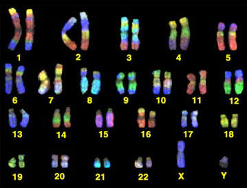
ChromosomesUnder normal conditions, humans have 23 pairs of chromosomes. Each chromosome is responsible for carrying genetic information in the form of DNA. The 23rd pair determines the gender, XX for a female and XY for a male. Chromosomes are inherited by the offspring during the process of meiosis, 23 individual chromosomes from each parent. All 3 billion bases that make up the human DNA can be found existing throughout all of the chromosomes. DNA has to be extremely wound up to make a chromosome. Histone proteins are the molecules that the DNA wraps around. When chromosomes are paired up, they are known as sister chromosomes, held together by a centromere. Each chromosome has a short arm (p) and a long arm (q). The chromosomes are located in the nucleus. Playing a key role in mitosis, their information is copied and transferred to a new cell. Chromosomes play in important role in the passing of the hemochromatosis disorder to offspring. This information is located in the meiosis section.

|

Hemochromatosis Genetic Basics
|
Hemochromatosis is located in the HFE gene, the short arm (p) of chromosome 6 (Cytogenetic Location: 6p22.2). This gene is responsible for the producing a protein that exists on the surfaces of cells, mostly liver or intestinal cells. This protein is responsible for detecting the amount of iron that is within the body. The HFE gene is also responsible for the creation and function of the hepcidin hormone, the largest iron regulatory hormone within the body. Hepcidin determines how much iron should be absorbed from foods as well as how much iron should be released from the body’s storage. Mutations C282Y, H63D, and S65C are three of the most common areas of mutation that exist on the HFE gene that can produce hemochromatosis.
|
|
